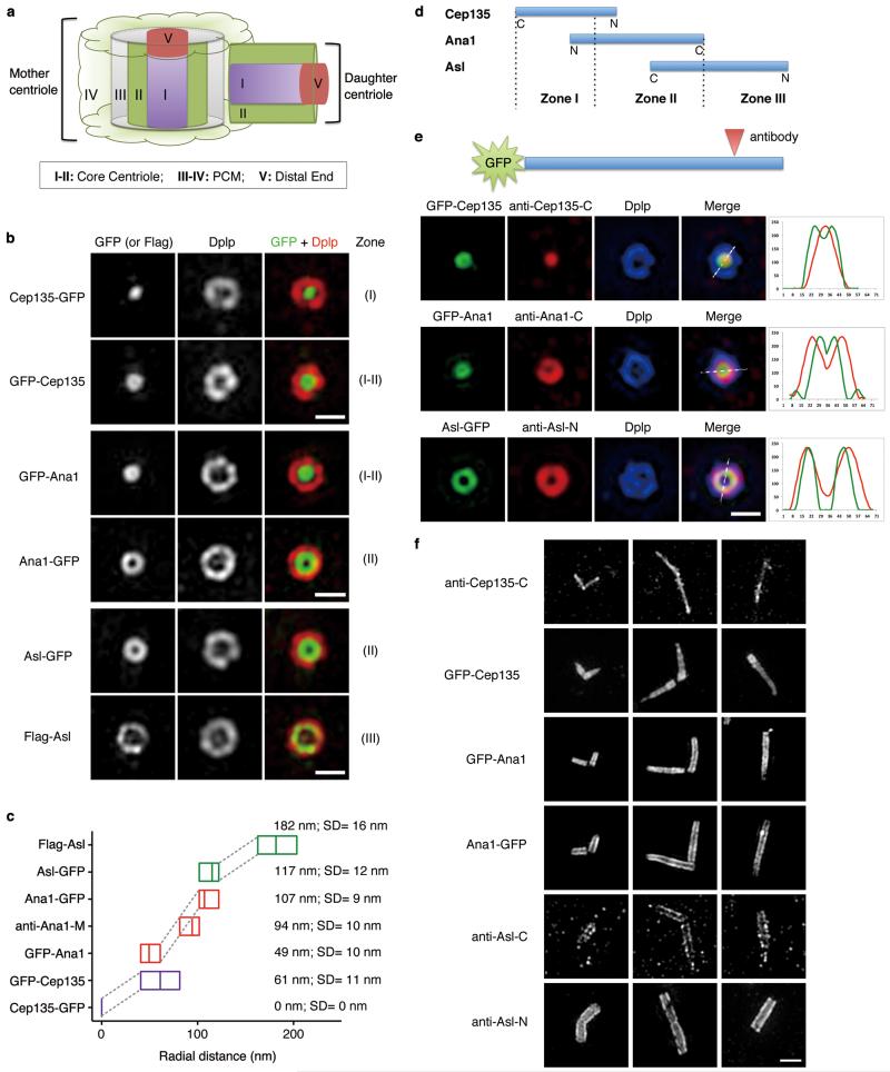
Figure 2. Cep135, Ana1 and Asl are extended molecules that span from inner to outer centriole.
(a) Scheme showing different Zones in Drosophila centrosome (modified from ref. 9).
(b) D.Mel-2 cells constitutively expressing GFP-tagged Cep135, Ana1 or Asl were immunostained to reveal Dplp and DNA (not shown). Cells expressing Flag-Asl were immunostained with additional anti-Flag antibody (bottom panel). The signal of Cep135, Ana1 or Asl appears in different Zones at centriole when the fluorophore-epitope is at different end of the protein. Scale bars, 500 nm.
(c) Average radial distance of different regions of Cep135, Ana1 or Asl. Low-high bar (horizontal) shows the range of radius and vertical line indicates mean. Cep135 in purple; Ana1, red; Asl, green. SD, Standard Deviation. From bottom up, n=35, 22, 14, 22, 15, 32 and 15 centrosomes, respectively.
(d) Map showing the relative positions of Cep135, Ana1 and Asl within single centriole. N and C indicate protein orientation.
(e) D.Mel-2 cells constitutively expressing GFP-tagged Cep135, Ana1 or Asl were immunostained for the respective antibodies recognizing the opposite end of the proteins and Dplp. Line scans reveal the stretched structure of exogenous Cep135, Ana1 and Asl at centriole. Scale bar, 500 nm.
(f) Representative images of centrioles in successive developmental stages of fly spermatocytes. Immunofluorescence signals are seen at positions of successively increasing diameter extending from (inner-most) C-terminus of Cep135; N-terminus of Cep135; N-terminus of Ana1; C-terminus of Ana1; C-terminus of Asl; to (outer-most) N-terminus of Asl. Scale bar, 1 μm.

