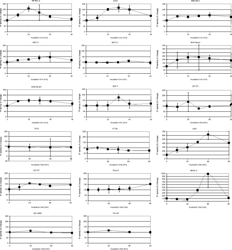
Fig. 3.

Time course of the microvesicle-associated TF activity released into the media by the cell lines. Cells (2 × 10⁵) were seeded out in 6-well plates and pre-adapted to respective serum-free medium. Microvesicle release was induced by incubation with PAR2-AP; SLIGRL; (20 μM). The released cell-derived microvesicles were isolated by ultracentrifugation at intervals up to 90 min and resuspended in Tris-saline. Microvesicle-associated TF activity was measured using the thrombin-generation assay, for each sample (n = 3)
