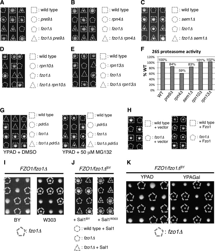
FIG 1.
Proteasome impairment restores growth of fzo1Δ cells. (A to E) Spores of heterozygous diploids were dissected on YPAD prior to growth at 26°C for 4 days. Genotypes of each colony are shown. (F) Suc-LLVY-MCA hydrolyzing activities of proteasome mutants used in panels A to E. Fluorescence intensities of cleaved MCA are normalized to that of wild-type cells. (G) Spores of the FZO1/fzo1Δ PDR5/pdr5Δ strain were dissected on YPAD containing dimethyl sulfoxide (DMSO) (vehicle, left panel) or proteasome inhibitor MG132 (right panel) prior to growth at 26°C for 4 days. (H) Spores of FZO1/fzo1Δ cells harboring empty vector or Fzo1-expressing plasmids were dissected on YPAD prior to growth at 26°C for 4 days. Genotypes of each colony are shown. (I) Spores of FZO1/fzo1Δ cells on the BY or W303 strain background were dissected on YPAD prior to growth at 26°C for 4 days. Cells lacking Fzo1 are indicated. (J) Spores of FZO1/fzo1ΔBY cells harboring Sal1BY- or Sal1W303-expressing plasmids were dissected on YPAD prior to growth at 26°C for 4 days. Genotypes of each colony are shown. (K) Spores of FZO1/fzo1ΔBY cells were dissected on YPAD or YPAGal prior to growth at 26°C for 4 days.

