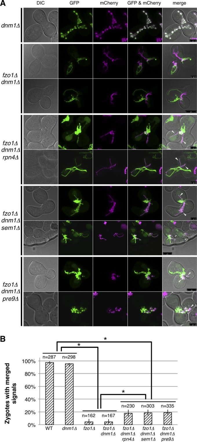
FIG 7.

Proteasome defects enhance Fzo1-independent mitochondrial fusion. (A) Cells expressing mitochondrially targeted GFP were mated to cells with mitochondrially targeted mCherry on YPAD and observed by confocal microscopy after 4 h. Images of DIC, GFP, mCherry, GFP/mCherry were merged as shown in the panels from left to right. (B) Percentage of cells with merged signals in mitochondrial fusion assay and 95% confidence intervals are shown. Counted cell numbers are indicated. Fisher's exact test was performed. Asterisks indicate P < 0.01.
