
FIG 3.
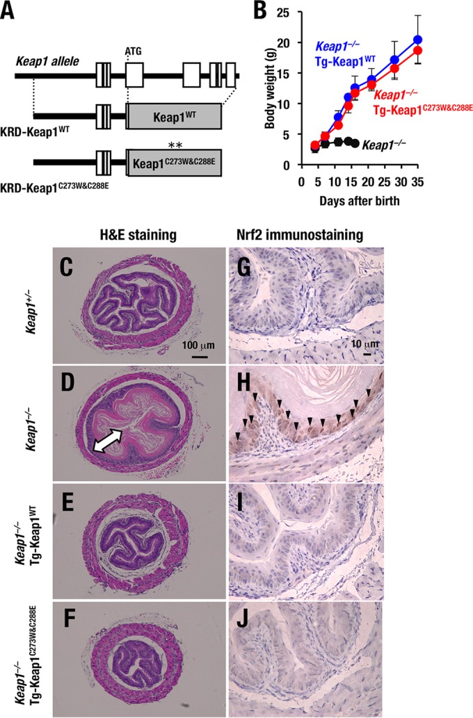
Keap1C273W&C288E represses Nrf2 activity in vivo. (A) A schematic presentation of KRD-Keap1C273W&C288E transgene that expresses Keap1 under the regulation of KRD is shown. (B) Growth curves for Keap1−/−, Keap1−/−::Tg-Keap1WT mice (line 34) and Keap1−/−::Tg-Keap1C273W&C288E mice (line 30). Note that mice of the last two genotypes grew normally. (C to F) Hematoxylin-eosin staining of esophagus transverse sections of Keap1+/− (C), Keap1−/− (D), Keap1−/−::Tg-Keap1WT (E), and Keap1−/−::Tg-Keap1C273W&C288E (F) mice at P10. The arrow in panel D indicates the thickened cornified layer. (G to J) Nrf2 immunostaining of esophagus transverse sections of Keap1+/− (G), Keap1−/− (H), Keap1−/−::Tg-Keap1WT (I), and Keap1−/−::Tg-Keap1C273W&C288E (J) mice at P10. Arrowheads indicate Nrf2 accumulation in basal layer cells of esophagi.