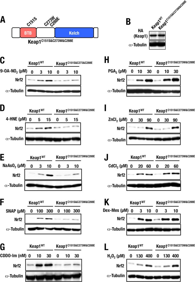
FIG 7.
Keap1 mutant with triple sensor cysteine mutations. (A) Schematic structure of Keap1C151S&C273W&C288E. (B) Keap1C151S&C273W&C288E protein levels of stable complemented MEFs examined by Western blotting. (C to L) Keap1WT and Keap1C151S&C273W&C288E MEFs treated with 0, 3, or 10 μM 9-OA-NO2 (C), 0, 5, or 15 μM 4-HNE (D), 0, 3, or 10 μM NaAsO2 (E), 0, 100, or 300 μM SNAP (F), 0, 10, or 30 nM CDDO-Im (G), 0, 10, or 30 μM PGA2 (H), 0, 30, or 90 μM ZnCl2 (I), 0, 20, or 60 μM CdCl2 (J), 0, 3, or 10 μM Dex-Mes (K), or 0, 130, or 400 μM H2O2 (L) for 3 h were examined by Western blotting.

