FIG 2.
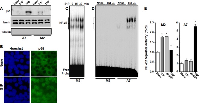
S1P promotes p65 translocation, NF-κB DNA binding, and reporter activity in FLNA-deficient M2 melanoma cells. (A) M2 and A7 cells were serum starved overnight and then stimulated with S1P (100 nM) or TNF (10 ng/ml) for 30 min. Nuclear fractions were prepared, and equal amounts of protein were analyzed by immunoblotting for p65. Lamin A/C was used as a nuclear marker, and tubulin was used as a cytosol marker. (B) M2 cells were serum starved overnight and then stimulated with S1P for 30 min. Cells were stained with Hoechst dye (blue) or p65 antibody (green) and visualized by confocal microscopy. Bar, 50 μm. (C and D) M2 and A7 cells were treated with S1P (100 nM) for the indicated times (C) or with TNF (10 ng/ml) for 30 min (D). NF-κB DNA binding activity in nuclear fractions (5 μg) was determined by EMSAs. (E) M2 and A7 cells were cotransfected with pNF-κB luciferase and β-galactosidase plasmids and then stimulated with S1P (100 nM), dihydro-S1P (dhS1P) (100 nM), or TNF (10 ng/ml) for 18 h. Luciferase activity was normalized to β-galactosidase activity and measured with the Dual-Light reporter gene assay. Data are expressed as fold increases and are means ± standard errors of the means from three independent experiments. *, P < 0.01 compared to no treatment.

