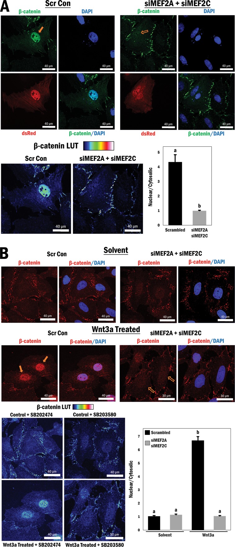
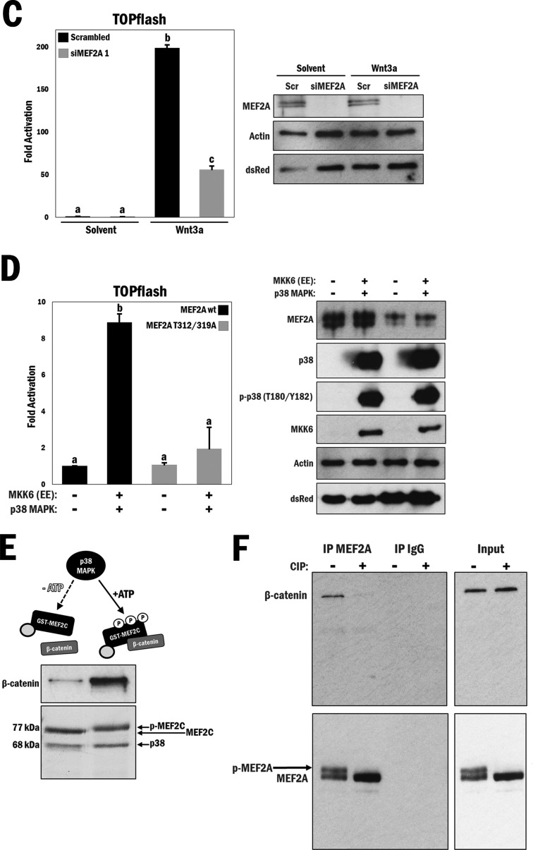
FIG 3.
MEF2 protein expression is required for β-catenin nuclear retention and enhanced by p38 MAPK-mediated phosphorylation of MEF2. (A) (Top) Cytomegalovirus (CMV)-driven dsRed was cotransfected with either scrambled siRNA (Scr Con) or siMEF2A/C RNA, followed by immunofluorescence staining of β-catenin (FITC) and the nucleus (DAPI), in A10 cells. (Bottom left) A rainbow LUT was used to indicate relative β-catenin intensities. (Bottom right) Relative nuclear/cytosolic fluorescence ratios were determined (n = 11 and 13, from left to right). b, P ≤ 0.0001. (B) (Top) Scrambled or siMEF2A/C RNA-transfected A10 cells underwent serum withdrawal for 12 h and were treated with either Wnt3a (200 ng/ml) or solvent (PBS; control) for 4 h, followed by immunofluorescence staining of β-catenin (TRITC) and the nucleus (DAPI). (Bottom left) A rainbow LUT was used to indicate relative β-catenin intensities. (Bottom right) Relative nuclear/cytosolic fluorescence ratios were determined (n = 28, 27, 27, and 28, from left to right). b, P ≤ 0.0001. (C) (Left) A10 cells were transfected with the TOPflash reporter and scrambled or siMEF2A RNA for 3 h and then cultured under serum conditions for 24 h, followed by treatment with either Wnt3a (200 ng/ml) or solvent (PBS) for 16 h in serum-free medium. b and c, P ≤ 0.0001. (Right) Corresponding Western blot analysis of cell lysates to confirm siRNA-mediated MEF2A depletion. (D) (Left) COS7 cells were transfected with the TOPflash reporter gene and different combinations of wild-type MEF2A, mutated MEF2A (T312/319A), MKK6 (EE), and p38 MAPK, as indicated. b, P ≤ 0.0001. (Right) Corresponding Western blot analysis. (E) Purified GST-MEF2C and GST-p38 MAPK were incubated at 30°C for 3 h, with or without ATP, as indicated in the figure, followed by addition of purified 6×His–β-catenin and GST-agarose beads for 1 h at room temperature. (Top) Immunoblot indicating relative amounts of β-catenin bound to MEF2C following precipitation. (Bottom) Representation of total MEF2C phosphorylation following 3 h of incubation with p38 MAPK and ATP. (F) A10 cell lysates (1 mg) were incubated with or without 10 U of CIP for 30 min at 37°C during immunoprecipitation using MEF2A antibody. Beads were then washed and analyzed for the corresponding β-catenin protein by Western blotting. Lysates without antibody-conjugated beads were similarly incubated as input samples.


