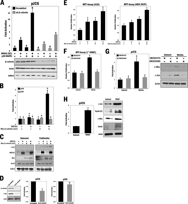
FIG 4.
MEF2 and β-catenin cooperate to promote cell proliferation in a p38 MAPK-dependent manner, and p38 MAPK regulates Wnt target gene protein levels. (A) (Top) c-Jun reporter gene (pJC6) activity was measured in A10 cells with various combinations of ectopically expressed MKK6 (EE) and p38 MAPK and either scrambled shRNA (control) or shRNA β-catenin, as indicated in the figure. b and c, P ≤ 0.0001. (Bottom) Corresponding Western blot analysis of cell lysates. (B) A10 cells were transfected with different combinations of MEF2C and Myc–β-catenin (act) and cotransfected with either pJC6 or pJSX (analogous promoter region containing mutations in the MEF2 binding site), as indicated. b, P ≤ 0.001. (C) Western blot analysis of either quiescent (serum withdrawal for 12 h) or proliferative (maintained with 10% FBS) A10 cells that were transfected with different combinations of MEF2C and Myc–β-catenin (act), as indicated. (D) Western blot analysis of A10 cells transfected with scrambled shRNA (control) or shRNA β-catenin, with comparative pJC6 and pJSX reporter activities. **, P ≤ 0.01; ns, not significant. (E) MTT cell proliferation assay in A10 (b, P ≤ 0.01) and HEK 293T (b and c, P ≤ 0.01) cells transfected with different combinations of MEF2C and Myc–β-catenin (act), as indicated. (F) MTT cell proliferation assay in primary VSMCs that underwent serum withdrawal (12 h) and were pretreated with SB203580 or SB202474 (10 μM) for 45 min and then stimulated with Wnt3a (200 ng/ml) or solvent (PBS) for 4 h. b, P ≤ 0.0001. (G) (Left) A10 cells were transfected with the pJC6 reporter for 3 h, followed by treatment with either Wnt3a (200 ng/ml), solvent (PBS), SB203580 (10 μM), or SB202474 (10 μM) for 16 h in serum-free medium, as indicated. b, P ≤ 0.001. (Right) Primary VSMCs were treated similarly to the manner described for panel F, and lysates were subjected to Western blot analysis as indicated. (H) (Left) pJC6 reporter assay in A10 cells transfected for 3 h and then treated with PDGF (20 ng/ml) or solvent (0.1% BSA in 4 mM HCl) for 16 h in serum-free medium. **, P ≤ 0.01. (Right) Western blot analysis of primary VSMCs that underwent serum withdrawal for 12 h and were treated with PDGF (20 ng/ml) or solvent (0.1% BSA in 4 mM HCl) for 4 h.

