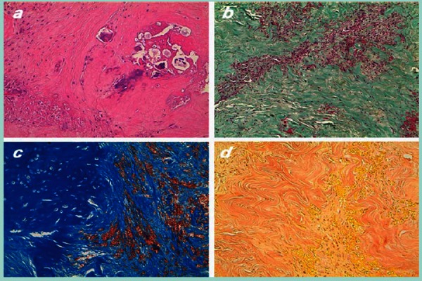
Figure 2.

Histological analysis of intraoperatory specimens with typical features of overload-related chronic tendonopathy. a) hematoxilin and eosin staining, showing condroid degeneration with calcic precipitates (20×); b) Goldner’s tricromie, showing dense collagen with reparative angioblastic tissue and haemorragic outflows (lO×); c) Azan Mallory staining, showing condromixoid degeneration with neo-formed vessels (20×); d) Van Gieson staining, showing angioblastic tissue and collagen dense bands (20×).
