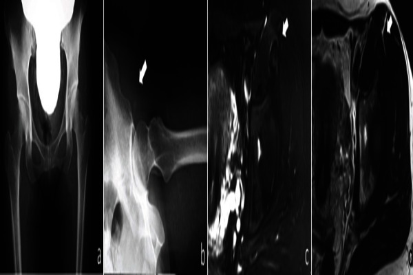
Figure 1.

(a) Standard anterior-posterior radiograph, (b, allow) X-ray of Lauenstein position is showing the calcification at the insertion of rectus femoris, (c) magnetic resonance image (MRI) of T2 fat suppression image shows high intensity change at the insertion of the rectus femoris, (d) MRI of T2 image.
