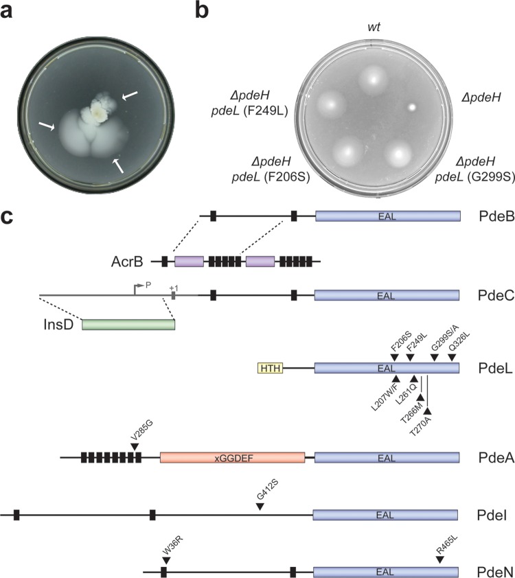
FIG 2.
Isolation of alleles activating E. coli phosphodiesterases. (a) Selection for motile suppressor mutants of a nonmotile ΔpdeH mutant strain on a low-percentage agar plate. Independent suppressors were recovered from motile flares (arrows) after incubation on motility plates for several days at 37°C. (b) Mutations in pdeL restore the motility of a ΔpdeH mutant. Mutant alleles of pdeL are indicated. Motility was examined as described for panel a. wt, wild type. (c) Graphical representation of isolated pde suppressor variants. Vertical black bars represent transmembrane helices, c-di-GMP-specific phosphodiesterase domains (EAL) are depicted in blue, the LuxR-like DNA binding domain of PdeL is shown in yellow (HTH), and the degenerate cyclase domain (xGGDEF) of PdeA is shown in red. The positions of single amino acid substitutions are marked with black triangles.

