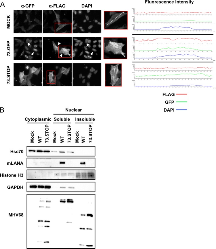
FIG 4.
mLANA enhances nuclear accumulation of Hsc70. (A) 3T12 fibroblasts transfected with plasmids encoding Hsc70-FLAG were mock infected or infected with 73.GFP or 73.STOP MHV68 at an MOI of 5 PFU/cell. Cells were fixed and permeabilized at 18 h postinfection and stained with antibodies specific for GFP and FLAG to visualize LANA and Hsc70 intracellular localization by fluorescence microscopy. DNA was detected with DAPI. White triangles designate infected cells exhibiting nuclear accumulation of Hsc70-FLAG. Fluorescence intensities for GFP, FLAG, and DAPI stains of the region marked within representative cells shown in the insets were plotted for each treatment. (B) 3T3 fibroblasts were mock infected or infected with WT MHV68 or 73.STOP MHV68 at an MOI of 5 PFU/cell. Cells were harvested 18 h postinfection, and cytoplasmic, soluble nuclear, and insoluble nuclear fractions were prepared. Equivalent amounts of protein for each fraction were resolved by SDS-PAGE, and immunoblot analyses were performed to detect the indicated proteins.

