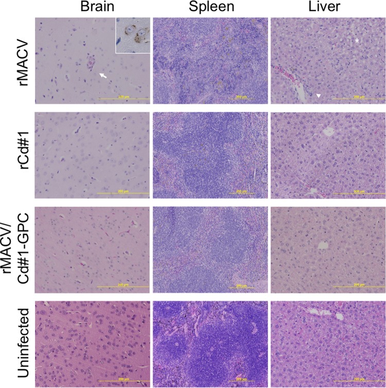
FIG 3.
Histopathological changes in the brain, spleen, and liver from infected mice at the terminal stage or 42 dpi. In the cerebrum, virus antigen-positive cells were broadly detected in rMACV-infected animals (inset at top left; magnification, ×40) but were not detected in rCd#1- and rMACV/Cd#1-GPC-infected animals. Endothelial hypertrophy (arrow) and vascular mononuclear infiltrates were observed in the brains of rMACV-infected animals. Microvesicular steatosis (asterisk) and perivascular mononuclear infiltrates (arrowhead) were present in the livers of rMACV-infected animals. Magnifications, ×20 (brain and liver) and ×10 (spleen).

