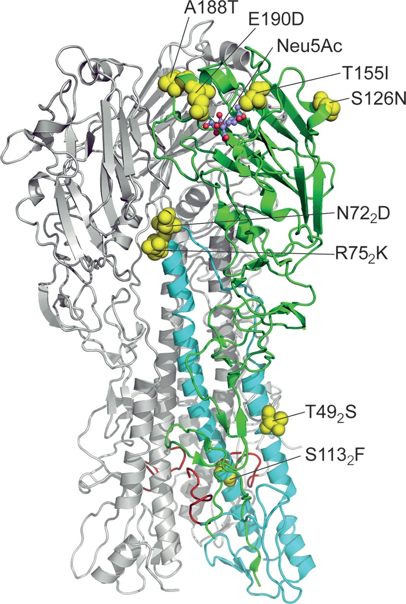
FIG 3.

Eight amino acid substitutions separating HAs of EAsw viruses from their putative avian ancestor. The model is based on the X-ray structure of the HA of A/duck/Alberta/35/1976 (2WRH, Protein Data Bank). For clarity, two HA monomers are colored gray, and the third monomer is colored green (HA1) and cyan (HA2). Location of substitutions is shown on this monomer as yellow spaced-filled models; sialic acid (Neu5Ac) in the receptor-binding site is shown as ball-and-stick model. Amino acids are numbered using H3 numbering system, with the subscript referring to HA2. Ten N-terminal amino acid residues of the fusion peptides of all three monomers are depicted in red.
