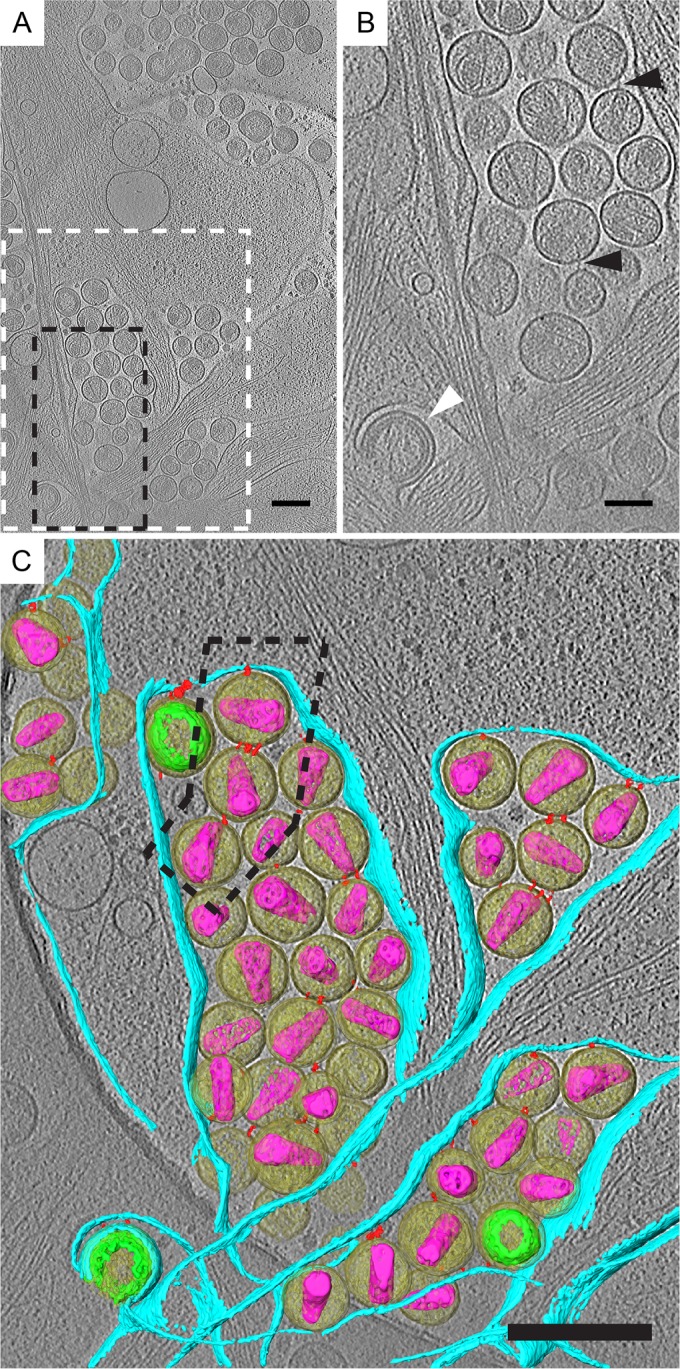
FIG 4.

Direct visualization of endogenous tethers on HIV-1 attached to HeLa cells by cryo-ET. HeLa cells transfected with pNLenv1ΔU were imaged by cryo-ET. (A) Tomographic slice (7.37 nm) of HIV-1 virions attached to plasma membrane. Immature and mature virions, maturation intermediates, and HIV budding from the plasma membrane were observed. (B) Enlarged tomographic slice. The dashed black box in panel A corresponds to panel B. Filamentous tethers attached to HIV-1 virions are indicated by black triangles. HIV-1 budding from the plasma membrane is indicated by the white triangle. (C) Segmented tomogram corresponding to the dashed white box in panel A. Elements shown include HIV-1 virions (yellow), immature Gag polyprotein (green), mature cores (purple), tethers (red), and the plasma membrane (cyan). Tethered HIV-1 elements are arranged as single chains, as indicated by the dashed black box around 3 HIV-1 virions connected to the cell. Scale bars are 200 (A and C) and 100 nm (B).
