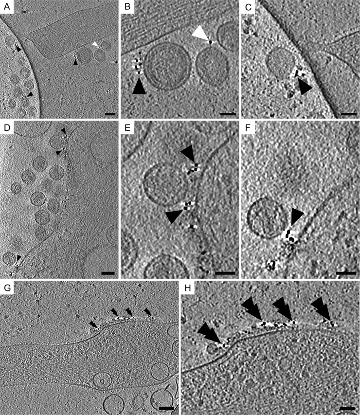
FIG 5.
Cryo-ET of native immunolabeled tetherin associated with HIV-1 VLPs and HIV-1 virions. Protein G conjugated to 6-nm colloidal gold distributed along the plasma membrane, on tethered HIV-1 VLPs, and on HIV-1 virions. (A) Tomographic slices (7.64 nm) of immunolabeled HT1080 cells transfected with plasmids encoding mCherry Gag (1:3 ratio of mCherry Gag and codon-optimized Gag) and pEGFP-tetherin. (B) Enlarged image of the dashed white box in panel A, which was magnified by a factor of 3. Shown is immunogold staining of tethered HIV-1 VLPs attached to the cell, as indicated by black triangles, and between other HIV-1 VLPs, as indicated by white triangles. (C) Enlarged image of the dashed black box in panel A, which was magnified by a factor of 3. (D) Tomographic slice (7.37 nm) of HIV-1 virions attached to the plasma membrane of an HT1080 cell (transfected with pNLenv1ΔU and pEGFP-tetherin). Black triangles point to immunogold staining. (E) Enlarged image of the dashed white box in panel D (magnified by a factor of 3). (F) Enlarged image of the dashed black box in panel D, which was magnified by a factor of 3. (G) Tomographic slice (7.64 nm) of a thin cellular extension devoid of HIV-1 virions but containing tetherin, as indicated by black double triangles. (H) Enlargement of area in panel G, which was magnified by a factor of 2. Scale bars are 100 nm (A, D, and G) and 50 nm (B, C, E, F, and H).

