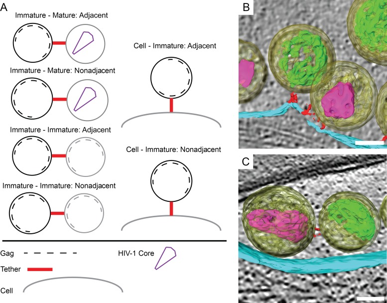
FIG 7.
Spatial relationship between tethers and the immature HIV-1 Gag lattice. (A) Tethers connected to immature HIV-1 virions were classified according to the arrangement of tethers relative to the Gag lattice. Tethers attached to an area of the viral membrane directly adjacent to the Gag lattice or nonadjacent to the Gag lattice were observed. (B) Example of an adjacent tether connecting an immature virion (yellow) to the plasma membrane (cyan). The tethers (red) are proximal to the Gag polyprotein (green). (C) Example of a nonadjacent tether adjoining an immature virion to a mature virion (mature core [purple]). The tether is making contact with the viral membrane in an area lacking an ordered immature Gag polyprotein. Scale bars are 50 nm.

