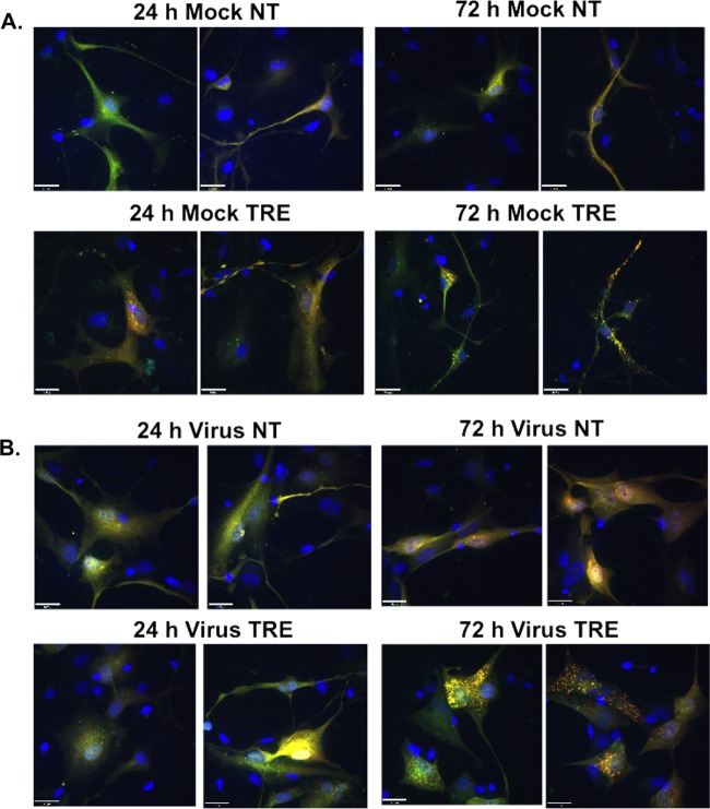
FIG 10.
Trehalose promotes acidification of autophagosomes in infected neuronal cultures. H9 day 8 neurons were transduced with a baculovirus expressing LC3B fused to GFP (green) and RFP (red). At 1 day postransduction, cells were mock infected (A) or infected with TB40E at an MOI of 3 (B) in the absence (not treated, NT) or presence (TRE) of 100 mM trehalose. At the indicated time points, cells were fixed, and nuclei were counterstained with Hoechst 33342. Z-stacks composed of individual 0.4-μm-thick slices were acquired at high magnification using a spinning-disk microscope. Between 8 and 10 fields were acquired for each condition. Representative phenotypes are shown. Colocalization (yellow) between the direct fluorescence of GFP and RFP results from a neutral pH, whereas red puncta correspond to a more acidic environment in which the GFP signal was quenched. Scale bar, 25 μm. Each experiment was repeated twice.

