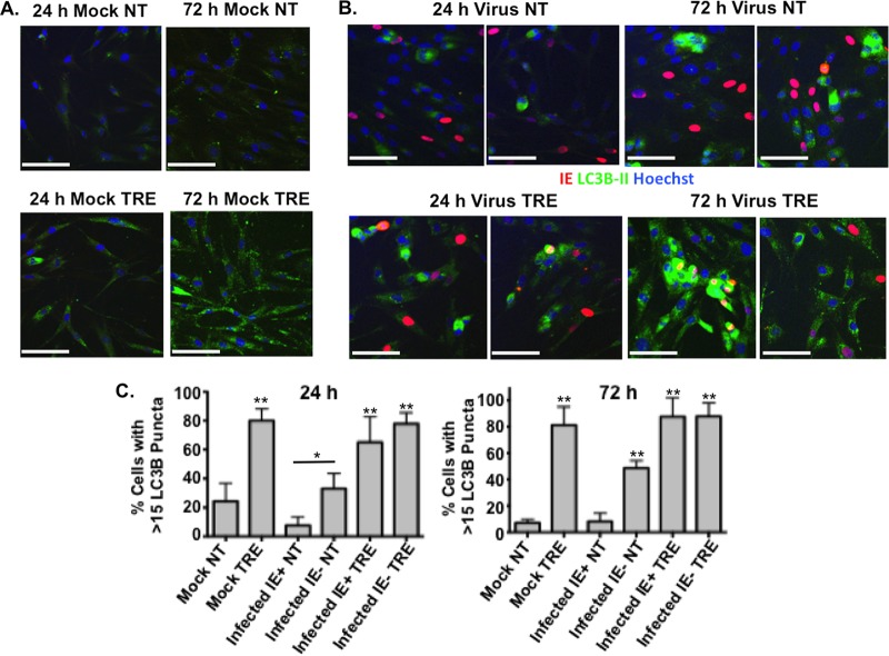
FIG 2.
Autophagosome formation in HCMV-infected cells is induced by trehalose. Synchronized HFFs were mock infected (A) or infected with TB40E at an MOI of 0.5 (B) upon seeding on coverslips in the absence (not treated, NT) or presence (TRE) of 100 mM trehalose. Cells were fixed at the indicated time points and permeabilized with saponin as described in Materials and Methods. Cells were stained with mouse monoclonal antibodies against LC3B (green) and IE1 (red). Nuclei were counterstained with Hoechst 33342 (blue). Pictures were acquired by confocal microscopy. Representative images are shown for each condition. Scale bar, 100 μm. (C) Quantification of the percentage of cells represented in panels A and B with more than 15 LC3B puncta. At least five fields (with approximately 25 cells in each) were counted for each condition. Histograms represent the means (bars), and the standard deviations (error bars) of the five fields are shown. Statistical significance was determined by one-way ANOVA combined with Tukey's multiple-comparison test (**, P < 0.01 versus results for mock uninfected and infected IE+ untreated cells; *, P < 0.05 versus results for infected IE+ untreated cells only). This experiment was repeated at least twice.

