FIG 5.
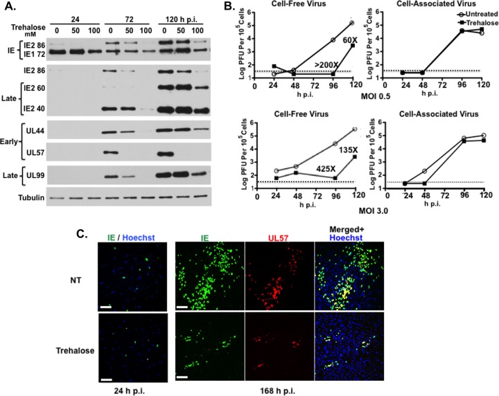
Trehalose inhibits HCMV gene expression, virus production, and viral spread in HFFs. (A) Synchronized HFFs were infected with TB40E at an MOI of 0.5 upon seeding in the absence or presence of 50 mM or 100 mM trehalose. Cell extracts prepared at 24, 72, and 120 hpi were analyzed by Western blotting using antibodies against HCMV IE1/IE2 (CH160), IE2-86, late IE2 proteins IE2-60 and IE2-40, UL44, UL57, and UL99. An antibody against α-tubulin was used to control for loading. The experiment was repeated three times, and a representative Western blot is shown. (B) Synchronized HFFs were infected with TB40E at an MOI of 0.5 or 3 upon seeding in the absence or presence of 100 mM trehalose. At the indicated time points, cell-associated and cell-free virus were prepared as described in Materials and Methods. Titers of viral preparations were determined by plaque assays. Graphs represent titer results obtained from a representative experiment. Relevant fold decreases in titers in trehalose-treated cells, relative to those nontreated cells, are indicated. Dotted lines represent assay limits of detection. (C) Synchronized HFFs were infected with TB40E at an MOI of 0.025 upon seeding on coverslips in the absence (not treated, NT) or presence of 100 mM trehalose. Cells were fixed at 24 and 168 hpi and stained with mouse monoclonal antibodies against HCMV IE (green) and UL57 (red) proteins. Nuclei were counterstained with Hoechst 33342 (blue). Images were acquired by epifluorescence microscopy. Representative images of phenotypes obtained at 24 and 168 hpi are shown. Scale bar, 100 μm. Each experiment was repeated three times.

