
FIG 4.
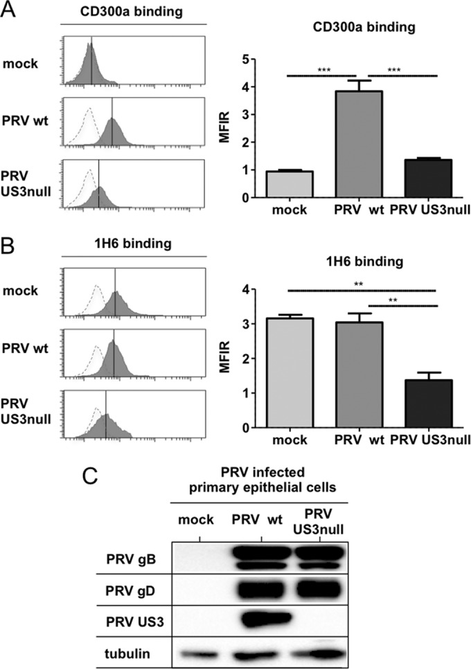
Modulation of CD300a binding and PS exposure by US3 also occurs in PRV-infected primary epithelial cells. Porcine primary epithelial cells were either mock infected or infected with WT or US3-null PRV (NIA3 strain) for 12 h. (A and B) Cells were assessed by flow cytometry for recombinant CD300a-Fc (1 μg/sample) binding (A) and PS exposure on the cell surface (by using antibody 1H6) (B). (Left) The x axes of histogram plots indicate fluorescence intensity, and vertical lines in histograms indicate median fluorescence intensity. Dotted-line histograms represent isotype-matched antibody control signals. (Right) Graphs show means + SEM for three independent repeats (**, P < 0.01; ***, P < 0.001). (C) Cells were assessed by Western blotting for the expression of PRV gB, PRV gD, PRV US3, and tubulin.