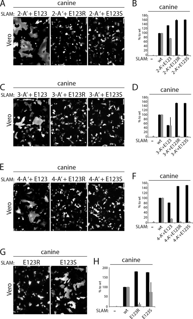
FIG 4.
Investigation of the impact of residue E123 of SLAM in triggering fusion. (A, C, E, and G) Cell-cell fusion activity in Vero cells triggered by coexpression of CDV H, CDV F, and cSLAM or a cSLAM mutant. To improve the sensitivity of the assay, the cells were additionally transfected with a plasmid encoding the red fluorescent protein. Images of fluorescence emissions from induced cell-cell fusion in representative fields are shown. The pictures were taken 24 h posttransfection with a confocal microscope (Fluoroview FV1000; Olympus). (B, D, F, and H) Dark-gray bars show the results for cell surface expression of the wt SLAM and SLAM mutants as determined by treating Vero cells 24 h posttransfection with an anti-HA MAb. After the addition of the secondary antibody, MFI values were recorded by flow cytometry. All values were normalized to the one recorded with the unmodified cSLAM molecule. Means ± SD of data from three independent experiments performed in triplicate are shown. Light-gray bars show the results for quantitative cell-cell fusion assay performed as described in the legend to Fig. 1F.

