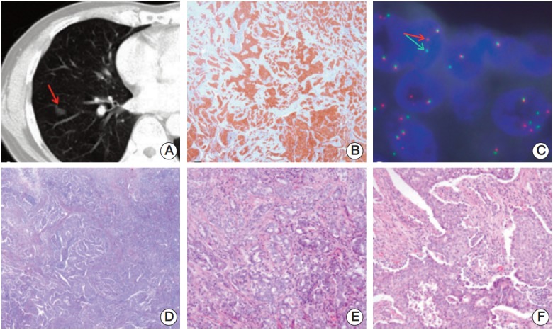
Fig. 1.

(A) Computed tomography scan of the chest showing a solid nodule with a speculated margin (arrow) measuring 1.5-cm in size in the lower lobe of the right lung. (B) Anaplastic lymphoma kinase (ALK) immunohistochemical staining showing cytoplasmic staining of tumor cells (×100). (C) Fluorescence in situ hybridization assay of ALK showing ALK genomic rearrangement by split 5'- and 3'-probe signals (arrows). (D-F) Hematoxylin and eosin staining showing adenocarcinoma (D, ×100), and a mixture of acinar pattern (E, ×200) and solid pattern (F, ×200).
