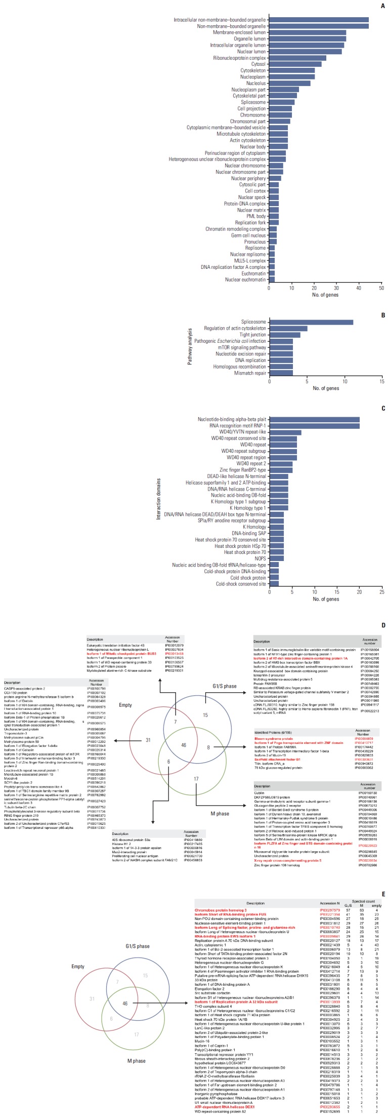
Fig. 2.

Proteins that interact with heterochromatin protein 1γ (HP1γ). (A) Diagram showing the cellular components of identified proteins that interact with HP1γ. (B) Diagram showing related molecular functions of these identified proteins. (C) Diagram showing domains that associate with HP1γ in identified proteins, visualized using Cytoscape. (D, E) Nonproportional Venn diagrams showing subsets of identified proteins in this study. Subset areas are not proportional to the actual relative subset sizes. Number of proteins identified in the immunoprecipitated complexes using the FLAG-(empty) vector-transfected cell lysates as a control (subset Empty), the FLAG-HP1γ vector-transfected cell lysates in the G1/S phase (subset G1/S), or the FLAG-HP1γ vector-transfected cell lysates in the M phase (subset M) are illustrated in the diagrams. The protein identities in each subset are described in the tables. In the subset tables. HP1γ-interacting protein that are implicated in DNA damage response pathways are marked in red.
