
Fig. 3.
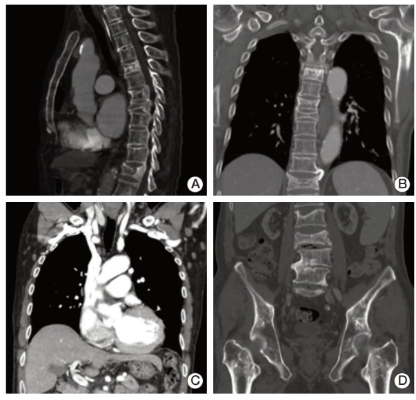
(A, B) Compression fractures were observed by a computed tomography scan of the patient’s chest at T5, T7-8, T12, and L3-4 spines. (C) In addition, computed tomography imaging identified cardiomegaly and multiple prominent lymph nodes in both axillae. (D) Irregular sclerotic change was observed in both femur necks.