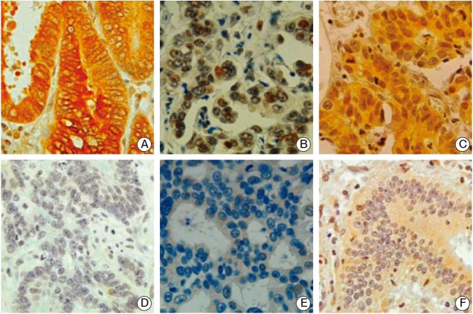
Fig. 4.
Representative immunohistochemical findings in human gastric cancer (GC) tissue specimens. (A) GC cells showing cytoplasmic phospho-FOXO1Ser256 (pFOXO1) expression with or without nuclear staining. (B) GC cells showing nuclear SIRT1 expression. (C) GC cells showing nuclear hypoxia inducible factor-1α (HIF-1α) expression with or without cytoplasmic staining. (D) GC cells without cytoplasmic pFOXO1 expression. (E) GC cells without nuclear SIRT1 expression. (F) GC cells without nuclear HIF-1α expression (A-F, ×400).

