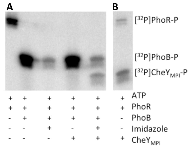
Figure 6.

The role of imidazole in an artificial phosphorelay. (A) Phosphorimaging scan of SDS-PAGE gel showing the presence of 32P on protein components in an artificial phosphorelay at pH 7.5. The PhoBN F20D variant was used for this analysis. (B) Phosphorimaging scan showing transfer of [32P] from PhoR to CheYMPI at pH 7.5. Reaction components in (A) and (B) were incubated for 30 minutes at room temperature.
