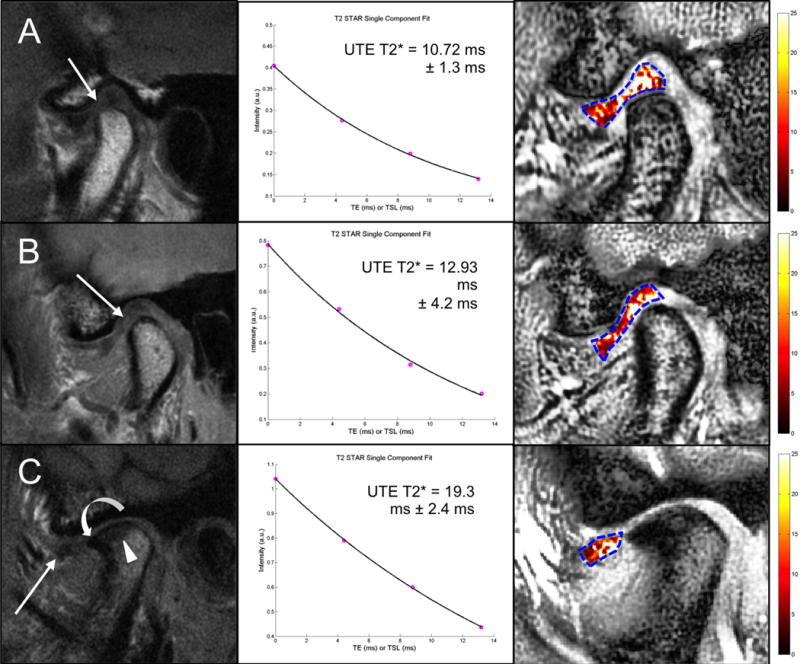
Figure 6.

Sagittal T1 NFS FSE MR images (left), UTE T2* mono-exponential fitting (center) and color mapping (right) of the articular disc of the TMJ in an asymptomatic subject (A), in a symptomatic subject with TMJ pain (B), and in a symptomatic subject who complained of pain and clicking in the right TMJ (C). A. The disc has a biconcave morphology with a focal area of hyperintensity in the intermediate zone and the anterior portion of the posterior band (arrow). B. The disc has a lengthened morphology with diffuse signal heterogeneity especially in the intermediate zone and the posterior band (arrow). C. There is anterior displacement of the articular disc, which appears small and irregular in shape with diffuse altered signal intensity especially to its posterior aspect (arrow). Associated findings in the mandibular condyle include thickening and irregularity of the subchondral bone plate and an osteophyte formation in the anterior margin of the condyle. In all subjects (A, B and C), color maps reflect the morphologic findings with increasing UTE T2* values at more advanced stages of disc alteration.
