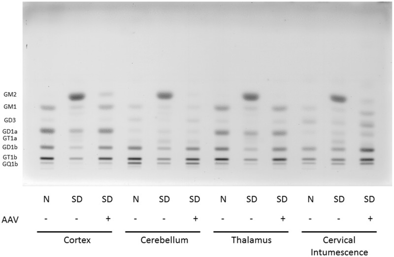
Figure 3.
HPTLC of gangliosides from Sandhoff cat cortex, cerebellum, thalamus, and spinal cord (cervical intumescence). Gangliosides from normal cats (N), Sandhoff disease cats (SD), and Sandhoff disease cats treated with AAV vectors were separated in a single ascending run with CHCl3:CH3OH;dH20, 55:45:10 (v/v/v) with 0.02% CaCl2. 1.5µg of sialic acid was spotted for each sample. The bands were visualized with the resorcinol-HCl spray. Ganglioside positions are shown on the left-hand side of the plate. AAV = adeno-associated virus; SD = Sandhoff disease; HPTLC = high-performance thin layer chromatography.

