Figure 1.
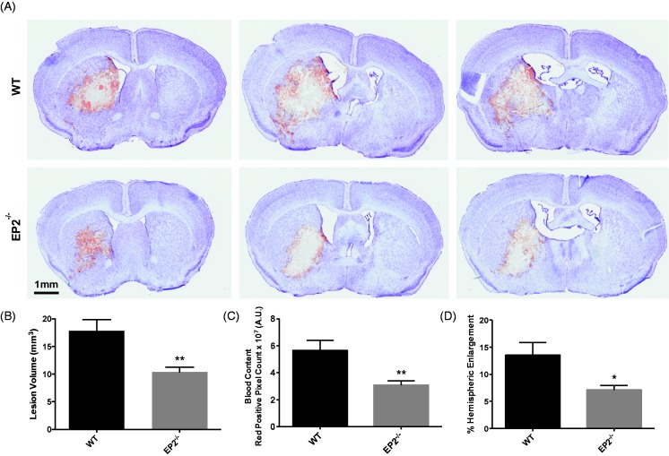
Genetic deletion of the PGE2 EP2 receptor reduces brain injury after ICH. WT and EP2−/− mice underwent ICH and were sacrificed at 72 hr for determination of lesion volume by Cresyl violet staining of brain sections. (a) Representative images of coronal brain sections from WT (upper panels) and EP2−/− mice (lower panels). Images are from the same animal and demonstrate characteristic hematoma profiles, where left to right corresponds to anterior to posterior. Center images are adjacent to the needle insertion site and represent maximal hematoma size. (b) Quantification of lesion volumes showed that EP2−/− mice had significantly less ICH-induced brain injury. (c) Red/brown positive pixel count analysis demonstrated that EP2−/− mice had significantly less blood accumulation within the injured brain area. (d) EP2−/− mice had less percentage of ipsilateral hemispheric enlargement. *p < .05 and **p < .01, all comparisons included n = 7 WT and n = 10 EP2−/− mice. PGE2 = prostaglandin E2; EP2 = E prostanoid receptor subtype 2; ICH = intracerebral hemorrhage; WT = wildtype.

