Figure 5.
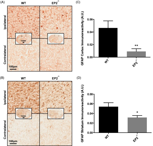
Effect of PGE2 EP2 receptor deletion on astrogliosis after ICH. Seventy-two hours after ICH, WT and EP2−/−mice were sacrificed and brains processed for GFAP immunohistochemistry to evaluate cortical and striatal astrogliosis. (a and b) Representative high magnification images of coronal brain sections showing the ipsilateral and contralateral (a) cortex and (b) striatum for WT (left panels) and EP2−/− mice (right panels). Square selections in the inserts denote magnified regions. (c and d) Quantification of brown positive pixel count demonstrated that EP2−/− mice had significantly less (c) cortical and (d) striatal GFAP immunoreactivity. Both groups demonstrated negligible staining in the contralateral cortex and striatum; thus, data are presented as ipsilateral immunoreactivity corrected for the area of quantification without normalization for the contralateral equivalent. Comparisons included n = 7 WT and n = 10 EP2−/− mice, *p < .05, **p < .01. PGE2 = prostaglandin E2; EP2 = E prostanoid receptor subtype 2; ICH = intracerebral hemorrhage; WT = wildtype; GFAP = glial fibrillary acidic protein.

