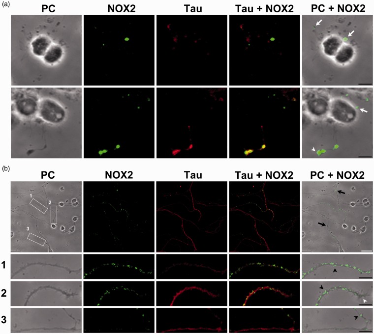
Figure 5.
NOX2 is expressed in filopodia and axonal growth cones in developing CGN. Representative confocal micrographs of NOX2 (green) and Tau (red) distribution and phase contrast (PC) micrographs at 0 and 3 DIV. (a) Two representative images of CGN at 0 DIV. White arrows indicate small protrusions and white arrowheads indicate growth cones. (b) A representative micrograph of CGN at 3 DIV. White squares (1 to 3) are shown below as magnified images. CGN were seeded at low density. White arrowheads indicate growth cones, black arrowheads indicate filopodia, and black arrows indicate varicosities (White scale bar, 20 µm; black scale bars, 5 µm). NOX = NADPH-oxidase; CGN = cerebellar granule neurons; DIV = days in vitro.

