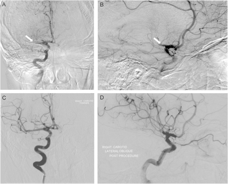
Fig. 2.
Successful intra-arterial treatment for an acute thromboembolic stroke involving the right middle cerebral artery (MCA). Pre-treatment digital subtraction angiogram—anterior (A) and lateral (B) views demonstrate proximal M1 occlusion of the right MCA. Defects are shown by arrows. Post-treatment—anterior (C) and lateral (D) views demonstrate recanalization of the proximal MCA and restoration of flow in its distal branches.

