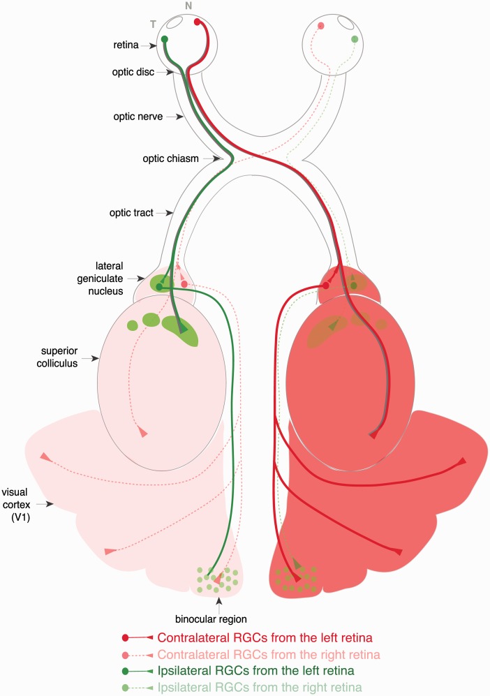
Figure 1.
Schematic representation of the visual pathway in the mouse. RGC axons transverse the eye to exit at the optic disk. The axons then travel via the optic nerves to the optic chiasm where they cross or avoid the midline to project ipsilaterally or contralaterally in the optic tracts to the main visual targets: the LGN in the thalamus or the superior colliculus. Ipsilateral and contralateral projections form distinct patterns at the visual nuclei. While ipsilateral axons form a few confined patches at the rostral LGN and SC (green spots), the contralateral terminals fill the rest of the tissue (red/pink). Second-order relay neurons transmit visual information from the thalamus to the visual cortex. The visual cortex of the mouse is mostly monocular, but there is a small binocular region that receives thalamocortical input from both hemispheres.

