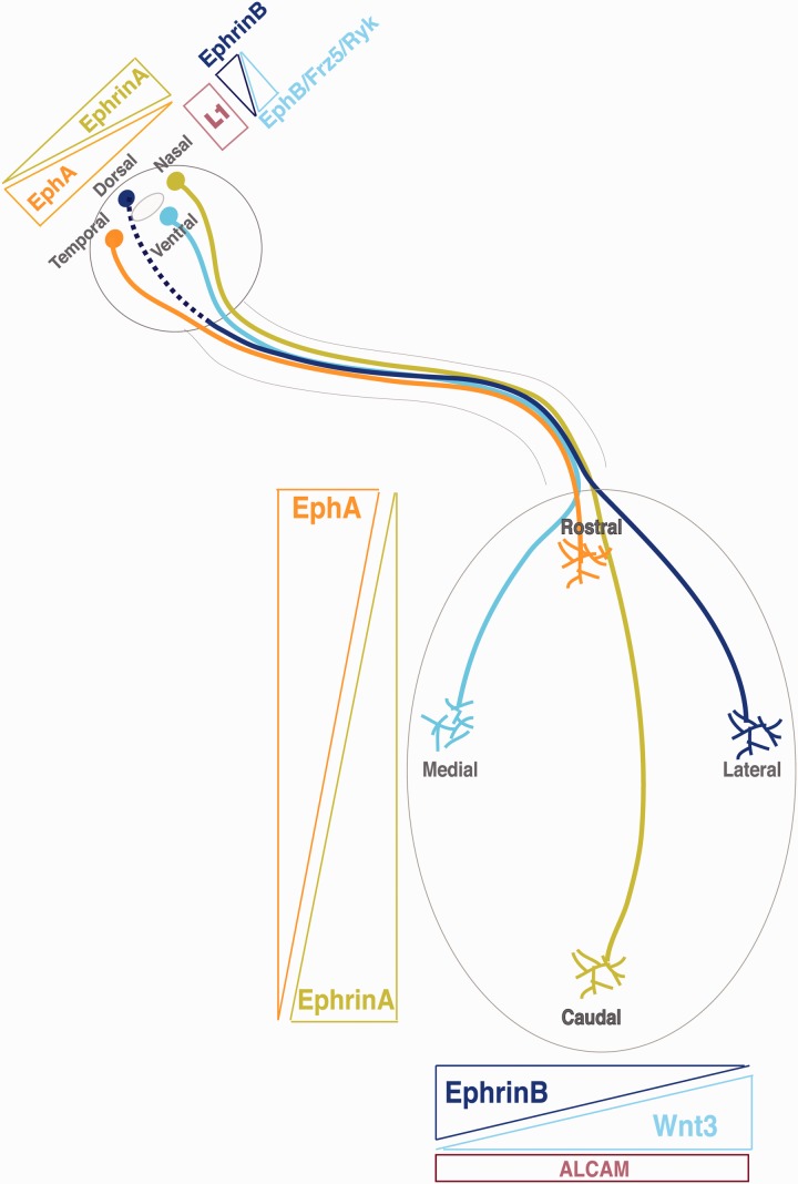
Figure 5.
Guidance molecules involved in topographic mapping at the superior colliculus of mice. The retinocollicular connection is highly organized, such that axons from neighboring neurons in the retina terminate in neighboring positions in the superior colliculus. Axons originating in the nasal retina terminate in the caudal SC, while temporal axons go to the rostral SC. In addition, axons originating in the dorsal retina map to the lateral SC and ventral axons terminate in the medial SC. Retinocollicular mapping is mediated by reciprocal molecular gradients in the retina and SC. EphA receptors are expressed by RGCs in an increasing nasal-temporal gradient. Neurons located more temporally express progressively more receptors. Ephrin-A ligands are expressed in the SC in an increasing rostro–caudal gradient. Nasal RGC axons project further into the SC because they express fewer EphA receptors and are subsequently less sensitive to the repulsive ephrin-A ligands. Conversely, the axons of temporal RGCs invade only a short distance into the SC because of their high sensitivity to ephrin-A ligands. In the dorsoventral axis, a gradient of EphB receptors exists in the retina with highest expression ventrally, while a gradient of ephrinB, highest medially, is expressed in the colliculus. In this case, however, EphB/ephrinB signaling seems to mediate attraction. Counterbalancing ephrinB1-EphB activity, a gradient of Wnt3 is highly expressed in the lateral SC and repels ventral axons expressing Ryk. Frizzled, also expressed by ventral axons, mediates attraction to medial SC cells expressing low levels of Wnt3. All RGC axons express the cell adhesion molecule L1-NCAM which, in combination with EphB/ephrinB signaling, seems to play a role in lateromedial mapping by interacting with ALCAM.

