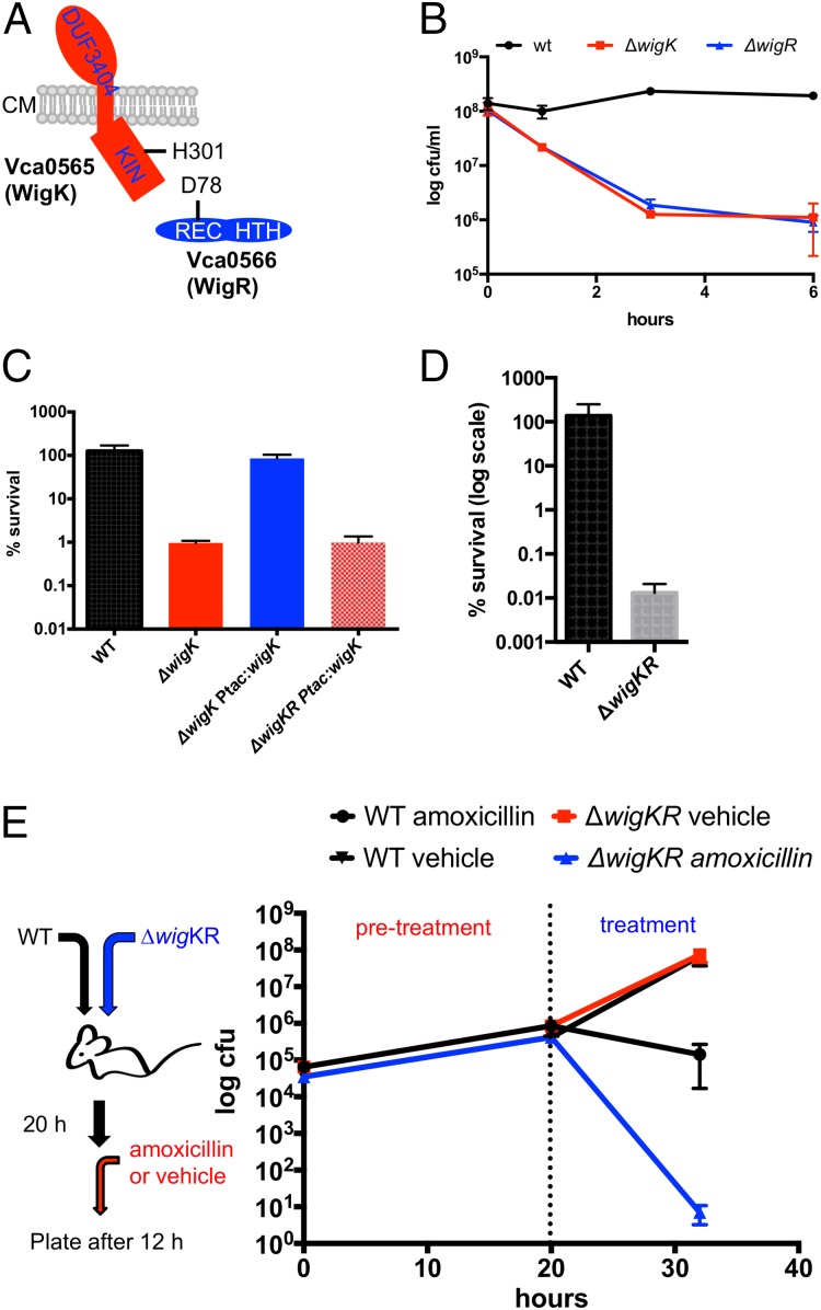
Fig. 1.
V. cholerae requires the wigKR locus to survive inhibition of cell wall synthesis. (A) Predicted domain organization of WigK and WigR showing their predicted sites of phosphorylation. Kin: kinase domain; REC: receiver domain; HTH: helix-turn-helix motif; CM: cytoplasmic membrane. (B) Survival of WT V. cholerae N16961 and its ∆wigK and ∆wigR derivatives after exposure to penicillin G [100 µg/mL, 20x minimum inhibitory concentration (MIC)] for the indicated amount of time. Data are averages of three biological replicates; error bars represent SE. (C) Complementation of wigKR-dependent penicillin sensitivity. Strains carrying complementing genes inserted in a single copy in a neutral chromosomal locus under an isopropyl β-d-1-thiogalactopyranoside (IPTG)-inducible promoter were grown to late exponential phase in LB + inducer and then exposed to 100 µg/mL penicillin G for 3 h; values represent mean of three biological replicates, and error bars represent SDs. (D) Survival of a ∆wigKR mutant in the presence of fosfomycin (200 µg/mL, ∼4× MIC). (E) WigKR are required for V. cholerae survival of beta-lactam treatment in vivo. N16961 (lacZ−) parental and ∆wigKR (lacZ+) were coinoculated into infant mice. Twenty hours after inoculation, mice were treated orally with either 50 mg/kg amoxicillin or vehicle control (“treatment”), and 12 h later cfu of the parental and ∆wigKR strains were determined. Data points are averages of 9 mice (amoxicillin) or 10 mice (vehicle); error bars represent SE.

