Fig. 1.
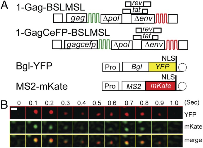
Detection of an HIV-1 RNA labeled with two different fluorescent proteins by using live-cell TIRF microscopy. (A) General structures of constructs used to detect HIV-1 RNA. HIV-1 constructs contain two sets of stem-loop sequences, BSL (green) and MSL (red), recognized by RNA-binding proteins BglG and MS2 coat protein, respectively. 1-GagCeFP-BSLMSL expresses a Gag-CeFP fusion protein, whereas 1-Gag-BSLMSL expresses an untagged Gag. Bgl-YFP and MS2-mKate are plasmids encoding fusions of RNA-binding proteins and fluorescent proteins. NLS, nuclear localization signal; Pro, RNA polymerase II promoter. Circle represents polyA signal. (B) A montage of selected frames from a movie showing simultaneous arrival and disappearance of YFP and mKate signals on the plasma membrane. (Upper) Signals detected in the YFP channel, (Middle) signals detected in the mKate channel, and (Lower) merged images of the YFP and mKate channels. Numbers on top indicate the imaged time in seconds. An LoG filter was applied by using ImageJ. (Scale bar, 1 μm.)

