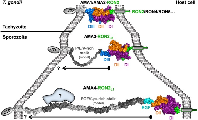
Fig. 5.
Variable stalk regions on AMA family members likely result in substantially different distances between the RON2-binding head group and the parasite cell membrane. (Top) TgAMA1 (purple/orange/blue)–TgRON2D3 (green) complex (PDB ID code 2Y8T). AMA1 TMD (generic seven-turn alpha-helix) is shown as a white surface. CTD, C-terminal domain. (Middle) TgAMA3–TgRON2L2D3 complex (PDB ID code 3ZLD) colored the same as in Top. Model of the Pro/Glu/Val rich stalk region is shown as a gray surface; the presented model represents a semiextended form, with the arrow reflecting the potential for considerable flexibility. (Bottom) TgAMA4–TgRON2L1D3 complex colored the same as in Top, with tandem EGF and Cys-rich domains connected head to tail and displayed as dark gray (EGF) and light gray (Cys-rich) surfaces. Blue shape with question mark indicates the possibility for the TgAMA4 stalk to recruit additional proteins. Arrow with question mark indicates potential for compaction.

