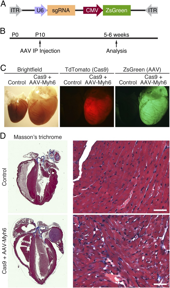
Fig. 2.
AAV9-driven expression of Myh6 sgRNA. (A) Myh6 sgRNA under the control of the U6 promoter was cloned into an AAV9 backbone, together with a CMV-driven ZsGreen reporter. (B) Animals were injected intraperitoneally at postnatal day 10 (P10) and subsequently analyzed 5–6 wk later. (C) An example of a Myh6-Cas9-2A-TdTomato heart (red, Center) that also received AAV-sgRNA against Myh6 exon 3 (green, Right). Compared with a littermate control animal, hearts from animals that received both Cas9 and sgRNA against Myh6 displayed extreme cardiac dilation and hypertrophy. (D) Histological section of a control heart and a heart that contained both Cas9 and AAV-sgRNA against Myh6 exon 3. Edited hearts displayed thinning of the ventricular walls and massive dilation of both the atria and ventricles. Minimal fibrosis was observed by Masson’s trichrome staining. (Scale bar, 40 μm.)

