Fig. 3.
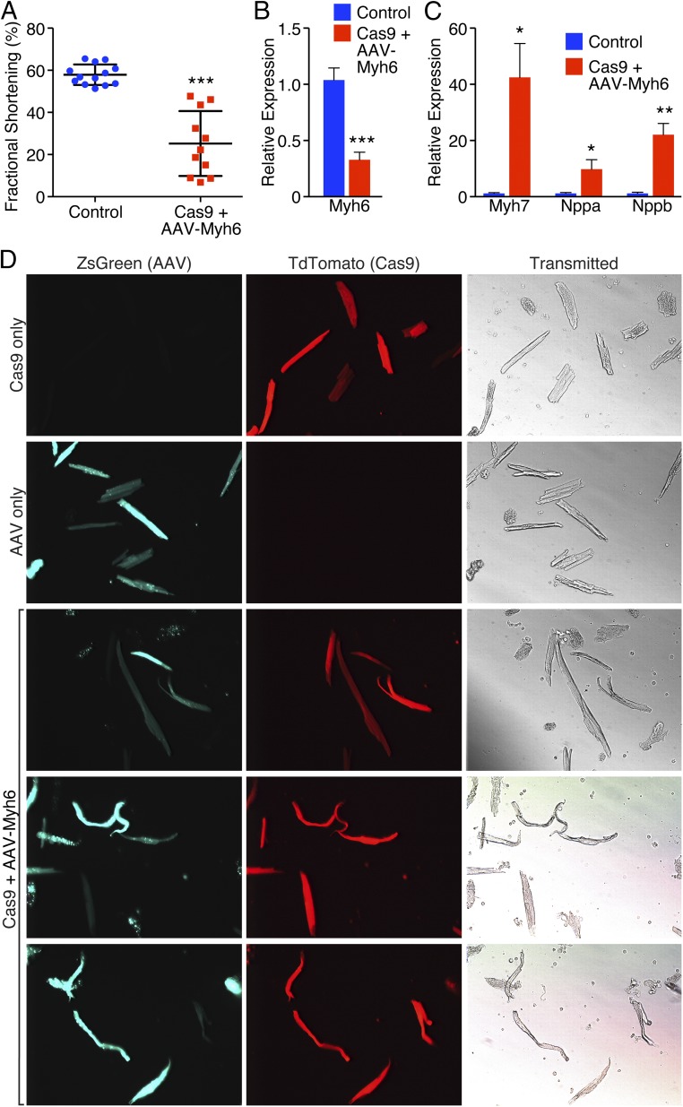
Cardioediting of Myh6 results in cardiac failure. (A) Echocardiography revealed a significant decrease in fractional shortening of animals that were Cas9+ and received AAV-sgRNA against Myh6 compared to littermate controls. ***P < 0.001; n = 13 control animals, 11 edited animals. (B) qPCR revealed that Myh6 expression was significantly down-regulated following knockdown of Myh6. ***P < 0.001; n = 5 control animals, 7 edited animals. (C) Myh7, Nppa, and Nppb were significantly up-regulated as detected by qPCR after Myh6 knockdown. *P < 0.05, **P < 0.01; n = 5 control animals, 7 edited animals. (D) Isolated cardiomyocytes from a (i) Myh6-Cas9-2A-TdTomato animal (top panel), (ii) wild-type animal that received AAV-sgRNA only (second panel), and (iii) Myh6-Cas9-2A-TdTomato positive animal that received AAV-sgRNA (bottom three panels). Edited animals display cardiomyocyte elongation characteristic of that seen in heart failure, as well as cardiomyocytes that are bent and floppy, suggesting loss of sarcomeric integrity and rigidity.

