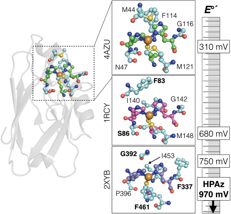
Fig. 1.
(Left) Tertiary structure of P. aeruginosa azurin, showing the location of the copper site. (Insets) Active sites of azurin (Top), Thiobacillus ferrooxidans rusticyanin (Middle), and Pycnoporus cinnabarinus laccase (Bottom). The invariant Cu-coordinating two His and one Cys are shown in green, magenta, and purple, respectively. The five residues mutated in HPAz, and those residues in the corresponding high-potential proteins, are shown in cyan. Bold lettering in the bottom two panels indicates residues that inspire the design of HPAz. The ruler shows the relative reduction potentials of each site. The Protein Data Bank codes are given to the left of each panel.

