
Fig 2. Effect of Tβ4 peptide on H2O2-induced cytotoxicity (A), Tβ4, inducible nitric oxide (NO) synthase (iNOS) and cyclooxygenase-2 (COX-2) mRNA and protein expressions (B, C), NO and prostaglandin E2 (PGE2) secretion (D, E) in PDLCs.
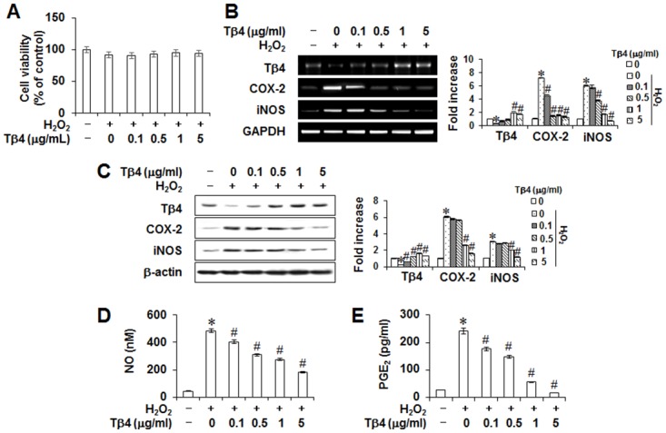
Cells were pretreated with indicated concentrations of Tβ4 peptide for 2 hours and then incubated with 200 μM H2O2 for 48 hours (A-E). Cell viability was measured by MTT assay (A). Protein and mRNA expressions were assessed by RT-PCR (B) and Western blot analysis (C), respectively. The production of NO (D) and PGE2 (E) were measured by Griess reaction and ELISA, respectively. Data replicated the quantifications of cytotoxicity, NO, and PGE2 with the standard deviation of at least three experiments (n = 4). The bar graph shows the fold increase in protein or mRNA expression compared with control cells. * Statistically significant differences compared with the control, p<0.05. # Statistically significant difference compared with the H2O2—treated group.