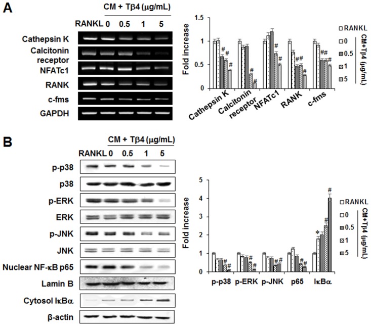
Fig 8. Effects of Tβ4 peptide on the functional changes of osteoclastogenesis (A) and MAPK and NF-κB signaling pathways in mouse BMMs.

Mouse BMMs were cultured with M-CSF (30 ng/mL) and RANKL (100 ng/mL) or CM collected from PDLCs for 5 days (A) and 60 minutes (B). The mRNAs expression was determined by PCR analysis (A). The phosphorylation of MAPKs (p38, JNK, and ERK), and activation of NF-κB were determined by Western blot analysis (B). Data were representative of three independent experiments. The bar graph shows the fold increase in protein or mRNA expression compared with control cells * Statistically significant differences compared with the control, p<0.05.
