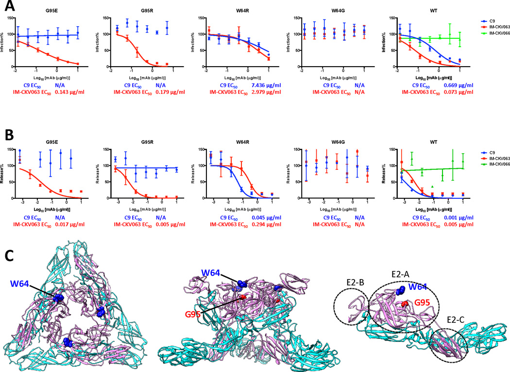
Figure 2. Mapping and characterize C9 and IM-CKV063 escape mutants.
(A) CHIKV-GLuc reporter viruses carrying indicated single escape mutations are resistant to antibody-mediated neutralization of entry (See also Figures S6). Similar to Figure 1B, WT and mutants GLuc reporter viruses were incubated with serially diluted mAbs before infecting RD cells at an MOI of 0.5. Infection was presented by GLuc activity in the supernatant at 24 hpi. Virus infection was normalized to a no antibody control. Results are representative of three independent experiments in triplicate. (B) CHIKV-GLuc reporter viruses encoding indicated single escape mutations are resistant to antibody-mediated inhibition of virus release (See also Figures S6). Similar to Figure 1C, RD cells were infected with WT or mutant GLuc reporter viruses for three hours before incubated with MAbs serially diluted in medium containing NH4Cl to prevent further rounds of infection. Virus release at 24 hours post-infection was measured by an infectivity assay. No antibody treatment was set to 100%. Data shown are representative of three experiments performed in triplicate with error bars representing standard deviation. (C) Mapping of C9 and IM-CKV063 escape residues on the crystal structure of the mature CHIKV envelope glycoprotein complex (PDB code 2XFB) in top view of trimer (left), side view of trimer (middle) and E1E2 dimer (right). Pink: E2; Cyan: E1.

