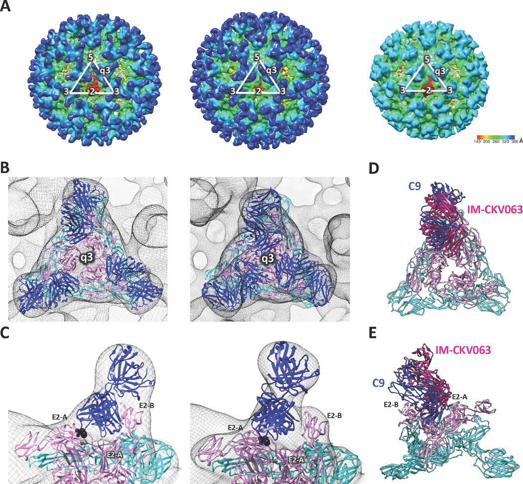
Figure 3. C9 and IM-CKV063 span two neighboring E2 molecules in one spike.
(A) CryoEM reconstruction of CHIKV (S27) virus like particle (VLP) (right), VLP in complex with C9 Fab fragments (left) and VLP in complex with IM-CKV063 fragments (middle). (B) Fitting of E1-E2 and Fab crystal structures into the viral spike at the q3-fold axis of the cryoEM density map of C9 Fab bound VLP (left) and IM-CKV063 Fab bound VLP (right). Blue, Fab; Pink, E2; Cyan, E1. (C) C9 Fab (left) and IM-CKV063 Fab (right) insert between domain A of one E2 (left) and domain B of a neighboring E2 (right) in one viral spike. E2-W64 was labeled in black and E2-G95 was labeled in gray. Comparison of C9 Fab (blue) and IM-CKV063 Fab (hot pink) binding to a viral spike in top view (D) and side view (E).

