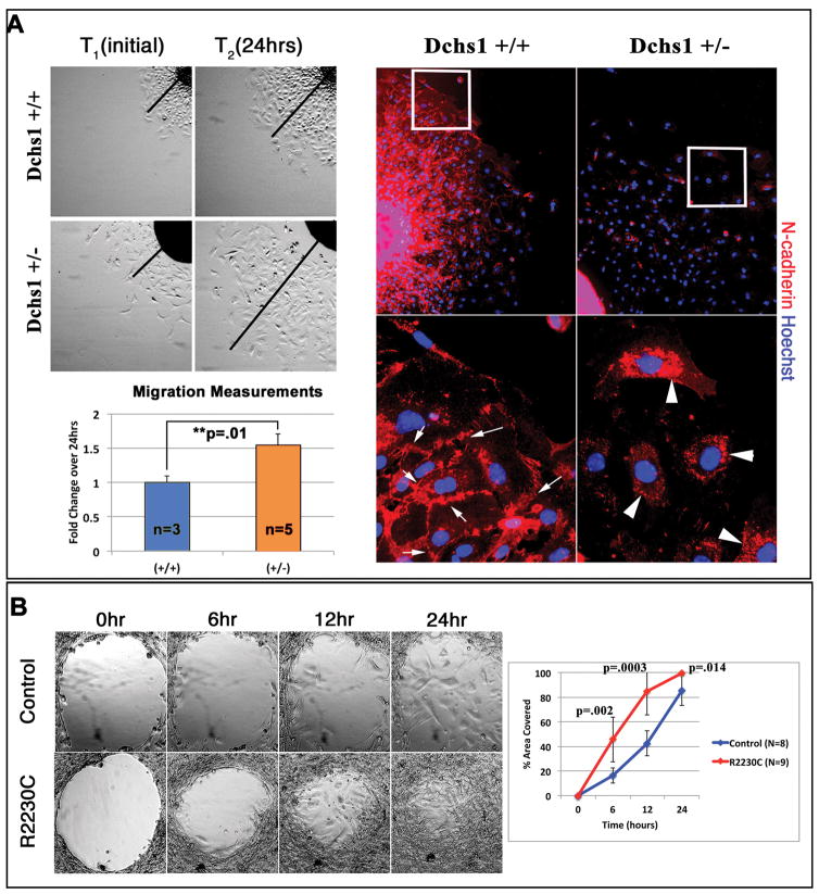
Extended Data Figure 10. Mice And MVP Patients With Dchs1 Deficiency Exhibit Migratory Defects In Vitro.
(A) Posterior Leaflets of P0 neonatal Dchs1+/+ and Dchs1+/− mice were explanted and interstitial cells were allowed to migrate out for 24 hours. Dchs1+/− mice exhibit increased migration (black lines drawn from explants) coincident with loss of cell-cell contacts and N-Cadherin expression at focal adhesions. Whereas N-Cadherin expression (red) is found at the membrane at points of cell-cell contract in Dchs1+/+ valvular interstitial cells (arrows), this membrane expression is lost in the Dchs1+/− cells and is prominently expressed in the cytoplasm (arrows). (Nuclei-blue). (B) Migration assays using control and MVP patient (p.R2330C) valvular interstitial cells exhibit a similar affect as observed in the mouse cells whereby the p.R2330C cells exhibit an increase in migration. p-values are denoted in graphs.

