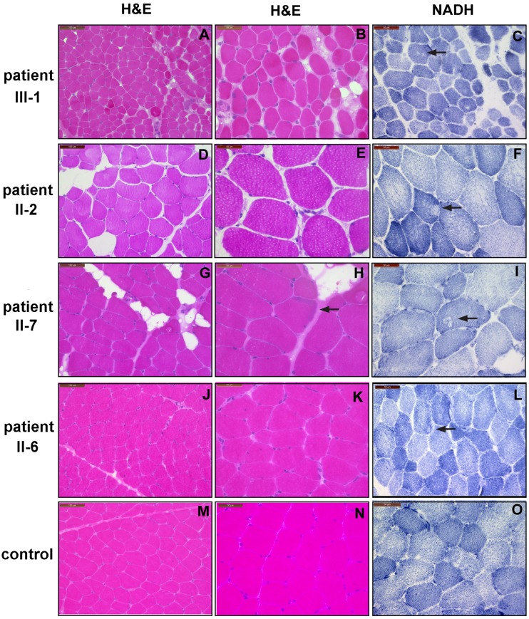
Fig 2. Histochemical and immunohistochemial biopsy analyses from affected family members and a normal control.
H&E staining of transverse cryostat sections displays findings well compatible with a slowly progressive myopathy with some replacement of muscle by fat in patients III-1 (A), II-2 (D), and II-7 (G); and is also abnormal in patient II-6 due to increased fiber variability (J) compared to a control (M). There is increased variability in fiber size in all family members (B,E,H,K) Notice also additional small atrophic fibers in patient II-2 (E), and some subsarcolemnal muscle fiber disorganization in patient II-7 (H arrow). NADH enzymehistochemistry depicts core like lesions in patients II-2 (F) and II-6 (I), and minor myofibrillar disorganization in patients III-1 (C) and II-6 (L).

