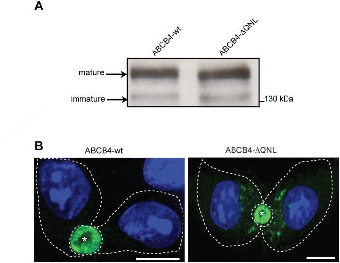
Fig 2. Expression and localization of ABCB4-wt and ABCB4-ΔQNL.
(A) ABCB4 was detected by immunoblotting from cell lysates of HepG2 cells stably expressing ABCB4-wt or ABCB4-ΔQNL. (B) HepG2 cells stably expressing ABCB4-wt or ABCB4-ΔQNL were fixed with methanol/acetone, processed for immunofluorescence using the monoclonal P3II-26 antibody and Alexa 488-conjugated anti-mouse IgG and visualized by confocal microscopy. Nuclei were stained with DRAQ 5 (blue). Asterisks indicate bile canaliculi. Cell contours are indicated by dotted lines. Bars, 10 μm. Representative of three experiments.

