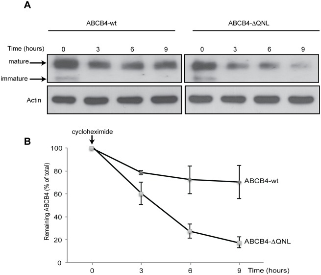
Fig 3. Stability of ABCB4-wt and ABCB4-ΔQNL.
(A) Stability of ABCB4-wt or ABCB4-ΔQNL was analyzed in stably transfected HepG2, after inhibiting protein synthesis with cycloheximide (25 μg/mL). ABCB4 was detected at the indicated time points in cell lysates by immunoblotting, using equal amounts of total proteins per lane. (B) Amounts of ABCB4 were quantified from chase experiments. The amount of ABCB4 at time zero was considered as 100%. Remaining ABCB4 at later time points was expressed as percentage of time zero. Means (± SEM) of three independent experiments are shown. * P<0.05 at all points.

